Research
How p53 structure becomes cellular decision
The Prives Laboratory studies the molecular logic of the p53 family from protein structure and DNA recognition to transcriptional networks, metabolism and tumour behaviour. The group examines how wild-type p53 is activated and restrained by MDM2 and MDMX, how p53 chooses among arrest, repair and death programmes, and how missense mutants accumulate and gain oncogenic activities. By combining biochemistry, structural and molecular biology, functional genomics, cell and organoid systems, and genetically defined cancer models, the laboratory aims to distinguish context-dependent p53 mechanisms and expose therapeutic vulnerabilities in p53-altered tumours.
01
Wild-type p53 structure and transcriptional choice
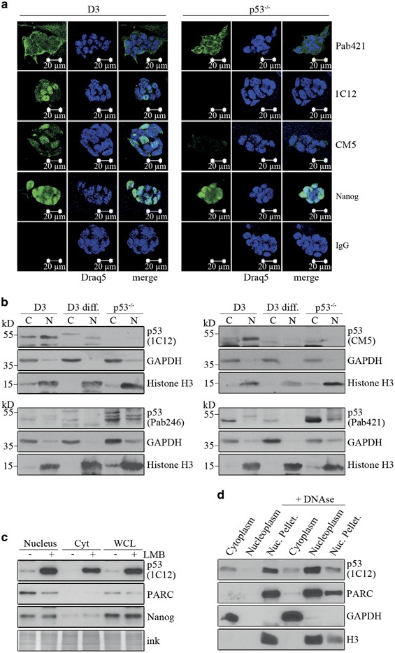
We define how p53 recognises chromatin, binds response elements and selects context-specific programmes of arrest, repair, metabolism and cell death.
Explore this programme →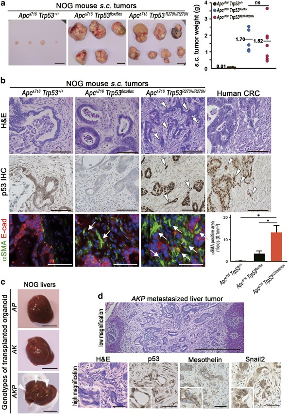
02
Mutant p53 gain of function and tumour evolution
We investigate when missense p53 mutants acquire oncogenic activities and how those activities depend on cellular lineage, cooperating pathways and tumour architecture.
Explore this programme →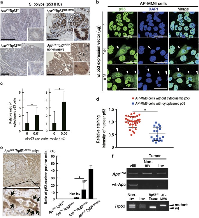
03
MDM2, MDMX and p53 protein stability
We study the regulatory circuitry that keeps wild-type p53 low in unstressed cells while permitting mutant p53 to accumulate in tumours.
Explore this programme →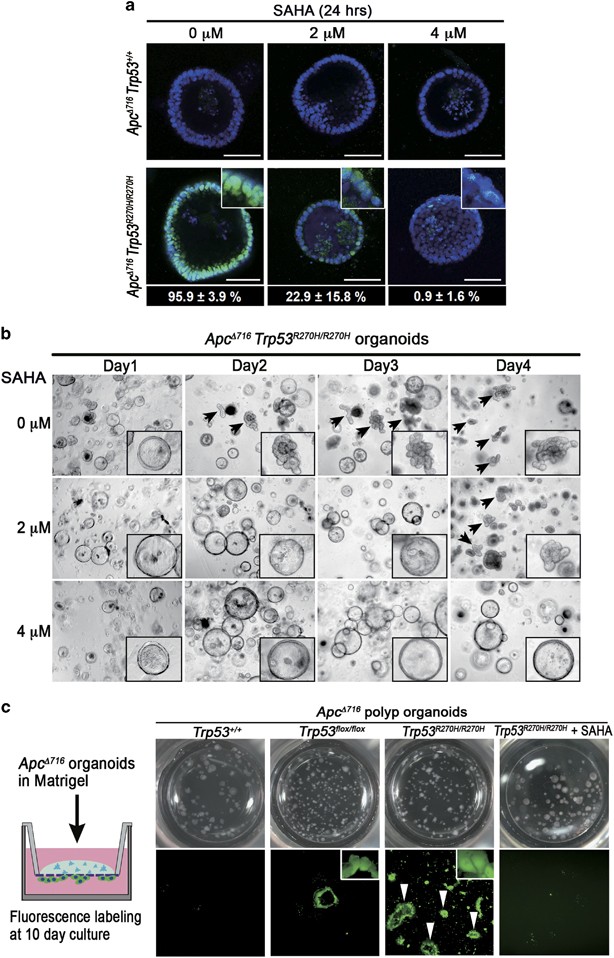
04
Metabolism, invasion and therapeutic vulnerabilities
We trace how p53 status rewires the mevalonate pathway, cytoskeletal signalling and tumour invasion to identify mutation-selective treatment opportunities.
Explore this programme →